

Shih Tzu Skin Cancer: Signs, Tumor Types, Diagnosis, and Treatment
Shih Tzu skin cancer can be difficult to recognize, especially when a small lump, unusual bump, or irritated patch of skin is hidden beneath your dog’s long, dense coat. Some skin tumors appear as harmless-looking warts or cysts, while others develop into firm masses, changing growths, or sores that refuse to heal. Although many lumps found on Shih Tzus are benign, certain malignant tumors, including mast cell tumors, melanomas, squamous cell carcinomas, and soft tissue sarcomas, require prompt veterinary attention. The challenge is that cancerous and noncancerous growths can look remarkably similar, making diagnosis from appearance or photographs unreliable. Understanding the warning signs, knowing how veterinarians examine skin growths, and recognizing when a lump needs testing can help you make informed decisions about your dog’s health. This guide explores the different types of skin tumors found in dogs, what research reveals about Shih Tzu cancer risk, the diagnostic procedures veterinarians use, available treatments, prognosis, and practical steps for monitoring your dog’s skin at home.
Table of Contents
- What to Know About Skin Tumors in Shih Tzus
- Types of Skin Tumors Found in Dogs
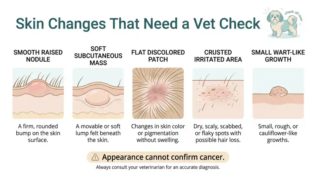
- Signs and Appearance of Skin Cancer in Shih Tzus
- Causes and Risk Factors
- When to Have a Shih Tzu’s Skin Lump Checked
- How Veterinarians Diagnose Skin Cancer
- Treatment Options for Shih Tzu Skin Cancer
- Recovery, Management, and Prognosis
- Reducing Risk and Monitoring Your Shih Tzu’s Skin
- Frequently Asked Questions
- Final Thoughts
What to Know About Skin Tumors in Shih Tzus
Skin tumors are abnormal growths that develop from cells within the skin or tissues immediately beneath it.
These growths may appear as raised bumps, thickened areas, discolored patches, or masses beneath the surface.
Some grow slowly and remain harmless. Others invade surrounding tissue or spread elsewhere in the body.
A Shih Tzu can develop skin tumors just like other dog breeds, particularly as it gets older.
However, discovering a skin lump does not automatically mean your dog has cancer.
The most important first step is determining what the growth actually is.
How Benign and Malignant Tumors Differ
A benign tumor is a noncancerous growth that generally remains localized and does not spread to distant organs.
Examples include many lipomas, sebaceous adenomas, and certain hair follicle tumors.
Benign tumors can still cause problems.
A large growth may interfere with walking, become irritated during grooming, or develop secondary infection.
A malignant tumor is cancerous. Its cells can invade surrounding tissues, and some malignant tumors can spread to other parts of the body.
This spread is called metastasis.
Not every malignant tumor behaves aggressively. Some remain localized for extended periods, while others grow rapidly or spread early.
| Feature | Benign Skin Tumor | Malignant Skin Tumor |
| Cancerous | No | Yes |
| Growth rate | Often slow, but variable | Slow or fast |
| Surrounding tissue invasion | Usually limited | May invade surrounding tissue |
| Distant spread | Does not metastasize | Some types can metastasize |
| Appearance | Variable | Variable |
| Veterinary diagnosis | Required to identify accurately | Required to confirm cancer |
| Treatment | Monitoring or removal | Depends on tumor type and stage |
A slowly growing lump is not necessarily benign, and a rapidly growing lump is not automatically malignant.
Veterinary testing is necessary to distinguish them reliably.
How Skin Tumors Differ from Allergies, Mange, and Other Skin Conditions
Shih Tzus can develop several skin conditions that resemble suspicious growths.
Common examples include atopic dermatitis, bacterial skin infections, yeast infections, and demodectic mange.
A skin allergy may cause itching, redness, inflammation, and hair loss.
Yeast infections can produce greasy skin, unpleasant odor, itching, and discoloration.
Demodectic mange results from excessive proliferation of Demodex mites and may cause patches of hair loss, scaling, and secondary infection.
These conditions can sometimes create swollen or thickened skin.
Meanwhile, certain cancers can resemble ordinary dermatitis.
For example, epitheliotropic lymphoma may initially resemble chronic inflammatory skin disease.
Shih Tzu skin cancer and yeast infection symptoms can therefore overlap, particularly when redness, itching, or abnormal skin texture occurs.
| Skin Condition | Possible Appearance | Important Distinction |
| Skin allergy | Redness, itching, inflamed patches | Often associated with recurrent itching |
| Yeast infection | Greasy, irritated, discolored skin | May have odor and inflammation |
| Demodectic mange | Hair loss, scaling, redness | Requires appropriate mite testing |
| Skin cyst | Rounded lump beneath or within skin | Contains material such as keratin |
| Benign tumor | Raised or underlying mass | Requires examination for identification |
| Skin cancer | Lump, plaque, sore, or abnormal patch | Can mimic several harmless conditions |
A veterinarian may use skin scrapings, cytology, cultures, or tissue samples to identify the cause.
Never assume a persistent growth is simply an allergy because your Shih Tzu has a history of sensitive skin.
What Research Says About Skin Tumor Risk in Shih Tzus
Studies have documented skin tumors in Shih Tzus, including both benign and malignant growths.
However, research findings must be interpreted carefully.
A South Korean retrospective study published in 2020 examined 2,172 canine skin tumor samples collected between 2005 and 2018.
Shih Tzus accounted for approximately 20% of the submitted tumor samples in that particular dataset.
Another retrospective study from Japan, covering 2008–2017, reported that Shih Tzus accounted for approximately 4.7% of its canine skin tumor cases.
These findings demonstrate that Shih Tzus are represented among dogs diagnosed with skin tumors.
They do not establish that 20% or 4.7% of all Shih Tzus develop skin cancer.
The studies examined submitted tumor samples, not every dog in the general population.
The number of Shih Tzus owned in a region, breed popularity, referral patterns, and veterinary testing practices can influence the results.
Additionally, the datasets include benign tumors, not just skin cancer.
Therefore, it would be misleading to conclude from these figures that Shih Tzus have a specific lifetime skin cancer risk.
How Age and Study Populations Affect Reported Breed Risk
Age is an important factor in canine skin tumors.
Many types become more common as dogs grow older, although younger dogs can also develop skin growths.
The Korean study described above reported a mean age of approximately 9.3 years among dogs with submitted skin tumor samples.
Other canine skin tumor studies have also reported substantial numbers of cases in middle-aged and senior dogs.
However, a tumor registry or biopsy database does not provide the same information as a population-based risk study.
A dataset may overrepresent dogs whose owners sought veterinary treatment or whose veterinarians submitted tissue for laboratory analysis.
It may also include repeated tumors from the same dog.
For individual Shih Tzu owners, the practical lesson is straightforward: age and breed history may help guide veterinary assessment, but they cannot establish whether a particular lump is dangerous.
Every new or changing growth deserves appropriate evaluation.
Types of Skin Tumors Found in Dogs
Several types of skin tumors can affect dogs, including Shih Tzus.
Some arise from skin cells, others from connective tissue, blood vessels, immune cells, pigment-producing cells, or skin glands.
Understanding the main categories helps explain why different tumors require different treatments.
Mast Cell Tumors
Mast cell tumors are among the most common malignant skin tumors diagnosed in dogs.
They develop from mast cells, immune-system cells involved in inflammatory and allergic responses.
Mast cells contain substances such as histamine and heparin.
When a mast cell tumor releases these substances, the surrounding tissue may become swollen, red, or irritated.
A mast cell tumor can resemble a harmless lump, wart, cyst, or fatty mass.
Some change size noticeably over a short period.
Possible signs include:
- A raised or underlying skin lump.
- Swelling that changes over time.
- Redness or bruised-looking skin.
- Itching or licking around the growth.
- Ulceration or bleeding.
- Occasionally, vomiting or other digestive symptoms.
Histamine release can contribute to gastrointestinal irritation and ulceration in some affected dogs.
Mast cell tumors are often evaluated initially using fine needle aspiration.
Tissue analysis is usually needed to establish tumor grade and support treatment planning.
Owners searching for mast cell Shih Tzu skin cancer pictures should remember that photographs cannot reliably distinguish these tumors from harmless growths.
A small, soft lump may still require testing.
Melanomas
Melanomas arise from melanocytes, the cells responsible for producing pigment.
These tumors can develop in the skin, mouth, nail bed, and other areas.
Some cutaneous melanocytic tumors in dogs are benign, while others are malignant.
Their behavior depends partly on location and microscopic characteristics.
A skin melanoma may appear as a dark brown or black mass.
However, not all melanomas contain obvious pigment, and some may look pink or pale.
The appearance alone cannot establish whether a melanocytic tumor is malignant.
Oral melanomas generally require particular attention because malignant oral melanoma can behave aggressively.
Owners concerned about Shih Tzu mouth cancer should understand that tumors inside the mouth are assessed separately from most skin tumors.
A veterinarian may recommend oral examination, biopsy, and additional imaging when a suspicious oral mass is present.
Squamous Cell Carcinomas
Squamous cell carcinoma, commonly shortened to SCC, develops from squamous epithelial cells.
These cells are found in the skin and other body surfaces.
Canine cutaneous SCC may appear as a firm growth, thickened plaque, crusted patch, or nonhealing ulcer.
Some lesions develop a wart-like surface.
Potential warning signs include persistent crusting, redness, ulceration, bleeding, and localized discomfort.
Certain squamous cell carcinomas are associated with prolonged ultraviolet radiation exposure, particularly on lightly pigmented or sparsely haired skin.
Others develop without an identifiable history of excessive sunlight.
SCC can also occur around the nail beds, where it may cause pain, swelling, or nail abnormalities.
The treatment and outlook depend on the location, tumor extent, and whether the disease has spread.
Fibrosarcomas and Other Soft Tissue Sarcomas
Fibrosarcoma is a malignant tumor that develops from fibrous connective tissue.
It belongs to a broader group called soft tissue sarcomas.
These tumors may originate from connective tissues beneath the skin.
A soft tissue sarcoma often appears as a firm or soft mass that gradually enlarges.
Some feel movable, while others become attached to surrounding tissues.
Many soft tissue sarcomas are locally invasive.
This means cancer cells may extend beyond the visible or palpable borders of the lump.
A mass can therefore appear relatively well defined while containing microscopic extensions.
Surgical removal is a common treatment, but planning the surgical margins carefully is important.
Some cases require radiation therapy or other treatments.
Hemangiosarcomas
Hemangiosarcoma is a malignant tumor that develops from cells associated with blood vessels.
In dogs, hemangiosarcoma can occur internally or involve the skin and underlying tissues.
Cutaneous hemangiosarcoma may appear as a red, dark, or bruised-looking growth.
It can sometimes bleed.
However, many noncancerous conditions can produce similar appearances.
The depth of the tumor matters.
Some superficial dermal hemangiosarcomas associated with sun exposure may have a better outlook after complete removal than tumors arising in deeper tissues.
Subcutaneous and internal forms can behave much more aggressively.
A veterinary pathologist must identify the tumor and its tissue involvement.
Epitheliotropic Lymphoma
Epitheliotropic lymphoma is an uncommon form of cutaneous lymphoma.
It involves malignant lymphocytes that affect the skin, often involving the epidermis and related structures.
This condition may initially look like chronic skin inflammation rather than a typical tumor.
Possible signs include:
- Persistent redness.
- Scaling or crusting.
- Hair loss.
- Thickened skin.
- Itching.
- Ulcerated areas.
- Multiple abnormal plaques or nodules.
Because these symptoms overlap with allergies and infections, diagnosis may be delayed.
A biopsy is generally required.
Treatment can involve medications, chemotherapy, and supportive care depending on disease extent.
Basal Cell Tumors and Carcinomas
Basal cell tumors are associated with cells in the skin’s basal layer or related follicular structures.
Terminology has changed over time, and some tumors previously described as basal cell tumors are now classified more specifically, including certain trichoblastomas.
Many of these growths in dogs are benign.
They may appear as small, firm, raised nodules, sometimes with hair loss over the surface.
Basal cell carcinoma is a malignant diagnosis and should not be confused with benign basal cell-type tumors.
Laboratory examination is important because the appearance may overlap with several other skin growths.
Sebaceous Gland Tumors
Sebaceous glands produce oily secretions that help lubricate the skin and coat.
Dogs can develop several types of sebaceous gland growths.
A sebaceous adenoma is a common benign tumor.
It may appear as a small, raised, irregular, or wart-like growth.
Sebaceous gland hyperplasia can create similar lesions.
Sebaceous epitheliomas have different biological characteristics, while sebaceous carcinomas are malignant but relatively uncommon.
These distinctions cannot be established simply by looking at the growth.
Owners sometimes search for “cancerous sebaceous cysts on Shih Tzu skin.”
However, a sebaceous cyst and a sebaceous gland tumor are not interchangeable diagnoses.
A true cyst is a sac-like structure containing material, whereas a tumor involves abnormal tissue growth.
A growth that resembles a cyst may require testing to determine what it is.
Hair Follicle Tumors
Hair follicle tumors arise from cells associated with hair follicles.
Examples include trichoblastomas, trichoepitheliomas, and pilomatricomas.
Many are benign, although malignant follicular tumors can occur.
These tumors may appear as raised skin nodules or lumps beneath the coat.
Some develop around the head, neck, or other body regions.
Because Shih Tzus have dense hair and require regular grooming, small follicular growths may be discovered during brushing or bathing.
Veterinary examination and tissue analysis help distinguish these tumors from cysts, inflammation, and other skin masses.
Histiocytic Cell Tumors
Histiocytic tumors arise from cells related to the immune system.
The most familiar benign example is a histiocytoma.
Histiocytomas often occur in younger dogs and may appear as small, raised, red, hairless, button-like growths.
Many resolve spontaneously, although veterinary examination is still important to confirm the likely diagnosis.
Histiocytic sarcoma is a different, malignant disease.
It should not be confused with a routine benign histiocytoma.
The distinction matters because these conditions have very different treatment and prognosis implications.
Benign Tumors, Cysts, Lipomas, and Papillomas
Many Shih Tzu skin lumps are not cancerous.
Common benign or non-neoplastic possibilities include:
Lipomas: Usually soft, fatty masses beneath the skin. They are commonly movable, although appearance and feel alone cannot confirm the diagnosis.
Skin cysts: Sac-like lesions containing material such as keratin.
Papillomas: Wart-like growths, some of which are associated with canine papillomavirus.
Skin tags: Small, often soft projections of skin.
Sebaceous adenomas: Benign growths associated with oil-producing glands.
Histiocytomas: Generally benign tumors, often seen in younger dogs.
Some benign growths can resemble malignant tumors, including mast cell tumors.
For this reason, a veterinarian may recommend fine needle aspiration or biopsy even when a lump appears harmless.
Signs and Appearance of Skin Cancer in Shih Tzus
The appearance of skin cancer varies considerably.
Some tumors form obvious lumps, while others produce flat patches, sores, thickened skin, or changes beneath the surface.
Recognizing these possibilities can help owners identify abnormalities early.
Lumps, Bumps, and Growths on or Under the Skin
A skin lump is one of the most common reasons owners seek veterinary evaluation.
The growth may feel soft, firm, rubbery, or irregular.
Some are attached to the skin, while others appear movable beneath it.
Potential locations include the back, chest, abdomen, legs, head, ears, paws, and tail region.
In Shih Tzus, long coat hair can make small growths difficult to see.
Owners may notice a lump only when bathing, grooming, or petting their dog.
Even a small growth deserves veterinary evaluation if it is new, unexplained, or changing.
Changes in a Lump’s Size, Shape, Color, or Texture
A previously stable lump that begins changing should be reassessed.
Watch for:
- Rapid enlargement.
- Changes in shape.
- New redness or discoloration.
- Increasing firmness.
- A previously movable lump becoming fixed.
- Repeated swelling and shrinking.
- The appearance of additional nearby growths.
Some mast cell tumors fluctuate in size because of inflammatory substances released from mast cells.
However, changing size is not unique to this type of cancer.
Inflammation, infection, and cysts may also change in appearance.
The pattern is useful information for the veterinarian, but not a diagnosis.
Sores, Bleeding, Ulceration, Itching, or Discharge
Skin cancer does not always look like a round lump.
It can appear as a persistent sore or abnormal skin patch.
Possible warning signs include:
- A wound that does not heal.
- Repeated bleeding.
- Crusting or ulceration.
- Unusual discharge.
- Persistent localized itching.
- Swelling around an abnormal patch.
- Darkening or loss of normal skin pigment.
- Skin thickening that continues to worsen.
These symptoms can also occur with infections, allergic dermatitis, and other conditions.
Nevertheless, persistent or worsening abnormalities should not be treated indefinitely at home without a diagnosis.
Owners searching for Shih Tzu skin cancer pictures should remember that two different diseases may look nearly identical in photographs.
Other Symptoms That May Occur If Cancer Spreads
Some malignant tumors remain localized, while others spread to lymph nodes or internal organs.
This process is called metastasis.
Depending on the tumor and affected organs, additional signs may include:
- Reduced appetite.
- Unexplained weight loss.
- Lethargy or weakness.
- Vomiting or diarrhea.
- Enlarged lymph nodes.
- Difficulty breathing.
- Changes in mobility.
- Persistent pain.
These symptoms are not specific to cancer.
For example, vomiting can occur with many digestive illnesses, and weakness may have several unrelated causes.
However, when a dog has a suspicious skin mass alongside significant changes in general health, prompt veterinary evaluation is important.
Severe breathing difficulty, collapse, uncontrolled bleeding, or marked weakness requires emergency care.
Causes and Risk Factors
The exact cause of many canine skin tumors remains unknown.
Cancer generally develops through genetic changes that affect normal cell growth and regulation.
Age, inherited susceptibility, environmental exposures, and other factors may influence the development of certain tumors.
Sun Exposure and Ultraviolet Radiation
Excessive ultraviolet radiation can damage skin cells.
In dogs, prolonged sun exposure has been associated with some cases of squamous cell carcinoma and superficial cutaneous hemangiosarcoma.
The risk is particularly relevant for lightly pigmented or sparsely haired skin.
A Shih Tzu’s long coat provides some physical protection, but it does not eliminate ultraviolet exposure.
Areas with thinner hair, such as the nose, belly, or sections affected by hair loss, may receive more direct sunlight.
Sun exposure is not responsible for every type of skin cancer.
Avoid assuming that a dog developed a tumor because it spent time outdoors.
Age and Genetic Factors
Cancer risk generally increases with age because cells accumulate changes over time.
Older dogs are more likely to develop several types of skin tumors.
However, young dogs can also develop skin masses, including benign histiocytomas and other growths.
Genetic factors may influence susceptibility to particular tumors in some breeds.
For Shih Tzus specifically, breed representation in tumor studies does not establish that every Shih Tzu has an inherited predisposition to skin cancer.
Owners should focus on individual health changes and regular veterinary examinations rather than relying solely on generalized breed-risk statements.
Viral Infections and Environmental Exposures
Some canine papillomas are associated with infection by canine papillomaviruses.
These viruses can cause wart-like growths, particularly in younger dogs.
Most ordinary canine papillomas are benign, although unusual lesions require veterinary evaluation.
Environmental factors may contribute to the development of certain cancers.
Examples include ultraviolet radiation and some chemical exposures.
However, for most individual canine skin tumors, a specific environmental cause cannot be identified.
It is not appropriate to assume that a household product, diet, or grooming treatment caused cancer without evidence.
How a Shih Tzu’s Coat Can Make Skin Changes Harder to Notice
The Shih Tzu’s long, dense coat is an important practical consideration.
Small skin lumps may remain hidden beneath the hair.
Areas of redness, discharge, or discoloration can also be difficult to identify without parting the coat.
Trapped moisture and debris can contribute to skin irritation or infection, but these conditions are not the same as cancer.
Regular grooming gives owners an opportunity to inspect the skin.
Pay particular attention to areas that are easy to overlook, including beneath the ears, around the armpits, along the abdomen, between the toes, and near the tail.
A new lump discovered during grooming should not be dismissed as a mat or skin tag without examination.
When to Have a Shih Tzu’s Skin Lump Checked
A useful rule is to arrange veterinary evaluation for any new, unexplained, or changing skin growth.
Not every lump requires emergency treatment, but waiting for a suspicious growth to become large or painful can delay important care.
Changes That Warrant a Prompt Veterinary Visit
Contact your veterinarian promptly if you notice:
- A new lump that has not been evaluated.
- A growth that enlarges rapidly.
- Bleeding or ulceration.
- A persistent nonhealing sore.
- Pain or significant swelling.
- A lump that changes color or texture.
- Repeated swelling and shrinking.
- A mass that interferes with walking, eating, or normal movement.
- A growth near the eye, mouth, or another sensitive location.
If your Shih Tzu also develops marked weakness, breathing difficulty, collapse, or uncontrolled bleeding, seek emergency veterinary care.
What Information and Photos to Share with the Veterinarian
Keeping a simple record can help your veterinarian understand how the growth has changed.
Useful information includes:
- When you first noticed the lump.
- Its location.
- Approximate size.
- Changes in color or texture.
- Whether it appears painful.
- Any bleeding or discharge.
- Whether your dog scratches or licks it.
- Previous lumps or tumor diagnoses.
- Changes in appetite, energy, or weight.
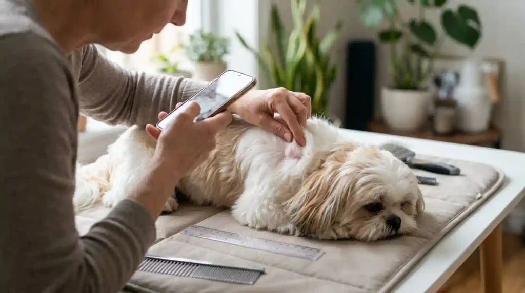
Take a clear, dated photograph with a ruler beside the growth when possible.
Photograph it under similar lighting conditions for future comparison.
Do not delay a veterinary appointment simply to collect multiple days of photographs.
Why Not to Squeeze, Cut, or Treat a Growth at Home
Trying to remove or treat an unexplained growth can cause injury and delay diagnosis.
Do not squeeze a suspected cyst or tumor.
Avoid attempting to cut off skin tags, puncture lumps, or apply human wart removers.
Some products can irritate or burn canine skin.
Manipulating certain tumors, particularly mast cell tumors, may also trigger localized swelling.
Home treatment may obscure the original appearance of the lesion and make examination more difficult.
If the area is irritated, prevent excessive licking and follow veterinary instructions for safe care.
Why Appearance Alone Cannot Identify Cancer
A veterinarian cannot reliably determine whether every skin mass is cancerous simply by looking at it.
Some malignant tumors resemble ordinary lipomas or cysts.
Some benign tumors look inflamed, irregular, or alarming.
The softness, color, or movement of a lump does not prove its biological behavior.
Similarly, internet photographs labeled “cancerous lump” cannot establish what is happening in your dog.
A veterinarian may recommend collecting cells or tissue for laboratory examination.
This is the most reliable way to move from suspicion toward an actual diagnosis.
How Veterinarians Diagnose Skin Cancer
Diagnosis usually begins with a physical examination and may involve collecting cells or tissue from the affected area.
The recommended tests depend on the mass’s location, appearance, size, and suspected tumor type.
Physical Examination and Skin Checks
The veterinarian will examine the growth and surrounding skin.
They may assess:
- Size and location.
- Texture and firmness.
- Mobility beneath the skin.
- Pain or inflammation.
- Nearby lymph nodes.
- Other skin abnormalities.
- General body condition.
The veterinarian may also search for additional lumps.
Some dogs have multiple unrelated skin growths, and each one may require separate assessment.
A growth that appears similar to a previously diagnosed benign mass should not automatically be assumed to have the same diagnosis.
Fine Needle Aspiration
Fine needle aspiration, often abbreviated as FNA, is a commonly used first-line diagnostic procedure.
A veterinarian inserts a small needle into the mass to collect cells.
The sample is placed onto a microscope slide for cytological examination.
This test is often quick and minimally invasive.
It can help identify certain tumors, including mast cell tumors and many lipomas.
However, FNA does not always provide a definitive diagnosis.
Some soft tissue sarcomas yield few cells, making the sample difficult to interpret.
A nondiagnostic result does not mean that the growth is harmless.
Further testing may be needed.
Biopsy and Laboratory Analysis
A biopsy involves collecting a piece of tissue or removing an entire growth for examination.
A veterinary pathologist studies the tissue under a microscope.
This process is called histopathology.
Histopathology can help identify the specific tumor type and, for certain cancers, provide information about biological behavior.
Two common approaches are:
Incisional biopsy: Removes a portion of a suspicious mass for diagnosis.
Excisional biopsy: Removes the entire mass, when appropriate, for diagnosis and potentially treatment.
The veterinarian selects the approach based on the tumor’s size, location, and suspected characteristics.
For some tumors, a planned biopsy before definitive surgery helps avoid inadequate removal.
Tumor Grade and Cancer Stage
Tumor grade and cancer stage describe different things.
Tumor grade refers to microscopic characteristics that help predict how aggressively certain cancer cells may behave.
Cancer stage describes the extent of the disease, including tumor size, local involvement, lymph nodes, and possible spread.
Not every skin tumor uses the same grading or staging system.
For example, mast cell tumors have specific grading approaches, while soft tissue sarcomas use other pathological criteria.
A veterinary oncologist may combine pathology results with examination findings and imaging to recommend treatment.
Blood Work and Imaging to Check for Spread
If a malignant tumor is diagnosed or strongly suspected, the veterinarian may recommend additional tests.
These can include:
- Complete blood count.
- Blood chemistry testing.
- Urinalysis.
- Lymph node sampling.
- Chest X-rays.
- Abdominal ultrasound.
- CT imaging in selected cases.
These tests may help assess overall health, guide anesthesia decisions, and investigate possible metastasis.
Not every skin lump requires every test.
The diagnostic plan should match the tumor type and the dog’s individual circumstances.
Treatment Options for Shih Tzu Skin Cancer
Treatment depends on the type of tumor, its location, size, grade, stage, and the dog’s overall health.
Some tumors can be treated successfully with surgery alone.
Others require additional therapy or long-term management.
Surgical Removal
Surgical removal is a common treatment for many localized skin cancers.
The veterinarian removes the tumor along with an appropriate amount of surrounding tissue.
The goal is to remove cancerous cells while protecting nearby structures.
Surgery may provide an excellent outcome for certain early, localized tumors.
However, not every mass can be removed with the same procedure.
Tumors near the eyes, toes, mouth, or other delicate areas may require specialized planning.
Referral to a veterinary surgeon may be recommended.
Understanding Surgical Margins
A surgical margin is the surrounding tissue removed along with the visible tumor.
Margins matter because some cancer cells extend microscopically beyond what can be seen or felt.
A pathologist examines the removed specimen to determine whether tumor cells are present near the cut edges.
Complete or clean margins generally indicate that tumor cells were not identified at the examined edges.
Incomplete or involved margins indicate that tumor cells reach one or more examined edges.
Even a report of clean margins must be interpreted with the type of cancer and adequacy of the surgical plan in mind.
Incomplete removal may increase the risk of local recurrence.
Depending on the tumor, additional surgery, radiation therapy, or monitoring may be recommended.
Chemotherapy
Chemotherapy uses medications to target cancer cells.
It may be recommended for certain aggressive tumors, cancers that have spread, or cases where surgery alone is unlikely to provide adequate control.
Not every dog with skin cancer needs chemotherapy.
Veterinary chemotherapy protocols are selected according to cancer type and treatment goals.
Potential side effects include nausea, vomiting, diarrhea, lethargy, appetite changes, and effects on blood cell counts.
Many dogs tolerate appropriately managed protocols reasonably well, but individual responses vary.
A veterinary oncologist can explain the expected benefits, possible side effects, and monitoring requirements.
Radiation Therapy
Radiation therapy uses targeted radiation to damage cancer cells.
It may be considered when a tumor cannot be fully removed or when surgery would affect an important body structure.
Radiation may also be used after surgery to reduce the risk of local recurrence in selected cases.
The treatment plan depends on the tumor’s characteristics and location.
Possible side effects include localized skin irritation, hair changes, and other effects related to the treated area.
Specialist evaluation is usually necessary.
Immunotherapy and Other Treatment Options
Some canine cancers may be treated with additional approaches beyond conventional surgery, chemotherapy, and radiation.
These may include targeted medications, selected immunotherapy approaches, or other specialist treatments.
For example, certain mast cell tumors may be eligible for targeted treatment depending on tumor characteristics and the dog’s circumstances.
Immunotherapy is not a universal treatment for canine skin cancer.
Other procedures, including cryotherapy and electrochemotherapy, may be considered for selected tumors in appropriate veterinary facilities.
Cryotherapy uses controlled freezing to destroy suitable abnormal tissue.
Electrochemotherapy combines chemotherapy medication with targeted electrical pulses to increase drug uptake into cells.
These treatments are not appropriate for every tumor.
A veterinary oncologist can explain which options have meaningful evidence for the specific diagnosis.
For further information about veterinary cancer diagnosis and specialist treatment, the Cornell University College of Veterinary Medicine’s Oncology Service provides an authoritative overview of canine cancer care.
Monitoring Some Benign Tumors
Not every benign tumor requires immediate removal.
A veterinarian may recommend monitoring a diagnosed lipoma or another harmless growth when it is small, stable, and not interfering with normal activity.
Monitoring should follow an appropriate diagnosis.
It is not the same as assuming an unexplained lump is benign.
A mass that grows, becomes painful, bleeds, or changes unexpectedly should be reassessed.
In some cases, removal is recommended even for benign growths because of location, discomfort, or repeated irritation.
Recovery, Management, and Prognosis
The outlook for a Shih Tzu with skin cancer depends heavily on the specific diagnosis.
Some tumors are treatable with excellent long-term results.
Others require ongoing management or have a more serious prognosis.
Follow-Up Visits and Monitoring for Recurrence
After treatment, your veterinarian may recommend scheduled rechecks.
These visits help monitor the surgical site, identify new skin masses, and assess possible recurrence.
Some cancers require repeated lymph node examinations or imaging.
The follow-up schedule depends on tumor type, grade, stage, and treatment response.
At home, continue checking the skin and documenting new changes.
A dog that has previously developed one tumor may later develop an unrelated growth.
Each new lesion should be evaluated rather than assumed to be the same tumor returning.
Managing Treatment Side Effects and Supporting Recovery
Recovery after surgery may involve temporary activity restrictions, wound care, and prescribed medication.
An Elizabethan collar or another protective device may be needed to prevent licking.
Follow your veterinarian’s instructions for cleaning, medication, feeding, and activity.
If your Shih Tzu undergoes chemotherapy or another cancer treatment, report concerning side effects promptly.
These may include:
- Persistent vomiting.
- Significant diarrhea.
- Refusal to eat.
- Marked weakness.
- Unusual bleeding.
- Fever or signs of illness.
- Increasing pain or swelling.
Do not give human pain medications or supplements without veterinary approval.
Some substances can interfere with treatment or be toxic to dogs.
How Tumor Type, Stage, and Location Affect Prognosis
Prognosis means the expected course or outcome of a disease.
There is no single survival estimate that applies to all cases of Shih Tzu skin cancer.
A small, completely removed, low-grade tumor may have an excellent outlook.
A high-grade malignancy that has spread may require extensive treatment and have a more guarded prognosis.
Important factors include:
- Tumor type.
- Tumor grade.
- Cancer stage.
- Size and depth.
- Location.
- Surgical margins.
- Whether metastasis has occurred.
- Overall health and age.
- Response to treatment.
Some dogs live for years following successful treatment.
Others experience recurrence or progression despite appropriate care.
When curative treatment is not possible, palliative care can focus on comfort, pain control, appetite, and quality of life.
Your veterinarian or veterinary oncologist can provide the most meaningful prognosis after diagnosis and staging.
Reducing Risk and Monitoring Your Shih Tzu’s Skin
Not every skin tumor can be prevented.
However, consistent grooming, skin observation, sensible sun protection, and veterinary care can help owners identify problems early and reduce certain avoidable exposures.
Checking the Skin Beneath the Coat During Grooming
Regular grooming provides an excellent opportunity to examine your Shih Tzu’s skin.
While brushing or bathing, gently part the coat and inspect the skin.
Pay attention to:
- Behind the ears.
- Around the neck and shoulders.
- Along the back.
- Beneath the chest and abdomen.
- Inside the armpits.
- Around the groin.
- Between the toes.
- Near the tail and rear end.
Use your fingers gently to feel for new lumps beneath the coat.
Do not squeeze suspicious growths.
If your dog has painful or matted areas, ask a veterinarian or professional groomer for assistance.
Keeping a Dated Photo and Change Log of Skin Growths
A simple skin-growth log can help you recognize changes.
Record the location, approximate size, and date the growth was discovered.
Include a photograph when possible.
For example:
| Date | Location | Observation | Action |
| October 15 | Left shoulder | Small, firm bump noticed | Book veterinary examination |
| October 18 | Left shoulder | No obvious size change | Share observation with veterinarian |
| After diagnosis | Left shoulder | Follow-up monitoring recommended | Follow veterinary schedule |
Photographs should complement, not replace, professional diagnosis.
Do not wait for a lump to change before seeking its initial veterinary assessment.

Limiting Excessive Sun Exposure
Reasonable sun protection can help reduce ultraviolet exposure to vulnerable skin.
Consider limiting prolonged outdoor exposure during periods of intense sunlight.
Provide shade during outdoor activities.
Dogs with lightly pigmented or sparsely haired areas may benefit from additional protection.
Your veterinarian can recommend an appropriate dog-safe sunscreen when needed.
Avoid applying human sunscreen without checking its ingredients and suitability, particularly products that may be harmful if licked.
Protective clothing may be useful for some dogs, provided it does not cause overheating or discomfort.
UV-blocking window screens or films may help reduce ultraviolet exposure in sunny indoor resting areas, although their benefits depend on the specific product and wavelength protection.
Remember that Shih Tzus can also be sensitive to heat because of their brachycephalic anatomy.
Avoid overheating during outdoor activities.
Sun protection can reduce certain exposures, but it cannot guarantee prevention of skin cancer.
When to Ask Your Veterinarian About Prevention
Ask your veterinarian about skin monitoring during routine wellness appointments.
This is particularly helpful for senior Shih Tzus, dogs with previous skin tumors, and dogs that repeatedly develop new growths.
Discuss whether any existing masses need measurement, sampling, or follow-up.
You can also ask about managing chronic skin irritation, maintaining the coat, and protecting sensitive areas from sunlight.
There is no proven supplement, diet, or home remedy that prevents every type of canine skin cancer.
The most useful approach is regular observation, timely testing, and treatment based on an accurate diagnosis.
Frequently Asked Questions
What Does Skin Cancer Look Like on a Shih Tzu?
Skin cancer may appear as a lump, changing bump, discolored patch, wart-like growth, or nonhealing sore, but appearance alone cannot confirm cancer.
Are All Skin Lumps in Shih Tzus Cancerous?
No, many lumps are benign cysts, lipomas, or other noncancerous growths, but veterinary examination is needed to identify them.
Is Skin Cancer Fatal for Dogs?
Not always; some skin cancers can be treated successfully, while aggressive or advanced tumors may become life-threatening.
How Long Can a Dog Live with Skin Cancer?
Survival varies greatly depending on tumor type, grade, stage, location, treatment, and the dog’s overall health.
Final Thoughts
Shih Tzu skin cancer can be difficult to recognize because malignant tumors often resemble harmless cysts, warts, or ordinary skin irritation. Your dog’s long coat may also hide small growths, making regular skin checks especially valuable. Although many Shih Tzu skin lumps are benign, new or changing masses deserve veterinary attention because appearance alone cannot determine whether cancer is present.
Early diagnosis through fine needle aspiration, biopsy, and other appropriate testing can help guide treatment decisions. Depending on the tumor, surgery, medication, radiation therapy, or specialist care may provide effective options.
The most important step is to have unexplained skin growths evaluated rather than waiting for them to become painful or visibly abnormal.
If you discover a new lump, take a dated photograph, record any changes, and schedule a veterinary appointment. Prompt professional care gives your Shih Tzu the best opportunity for appropriate treatment and long-term comfort.